Tibialis Posterior Tendinosis
What is the Tibialis Posterior Tendon?
The tibialis posterior tendon is a strong tendon that arises from a large muscle deep in the calf, runs behind the inside of the ankle and attaches to a bone at the top of the arch of the foot( Navicular bone). The tendon is very important, as not only does it hold the arch of the foot up and keep the heel in-line, it also acts to stiffen the foot, turning it into a solid lever which allows the strong Achilles to push off with great power.
What is Tibialis Posterior tendinosis?
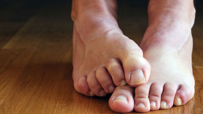
Tibialis posterior tendinosis is the most common cause of ‘Adult Acquired Flat Foot’ , it is a degenerative condition of the tendon, which if untreated leads to progressive weakening. Initially the tendon is still ‘tight’ and functions normally the foot remains normally shaped, but the inside of the ankle is painful; with time the tendon becomes ‘slack’ and weak the foot becomes flatter and the heel tilts outwards, pain may be experienced at the outside of the ankle, walking can become very tiring. Eventually the joints around the heel become stiff, painful and arthritic.
It is most common in females over 40 years of age, sportsmen and women are another group who may be affected.
What causes Tibialis Posterior Tendinosis?
Tibialis Posterior tendinosis probably results from a number of factors, related to the blood supply to the tendon, age related change to the collagen fibres that make up the tendon, injury and possibly pre-existing foot deformity. Most commonly however symptoms occur without an obvious precipitating event.
A small proportion of patients with tibialis posterior tendinosis have a slightly different tendon anatomy, having an extra bone, called an ‘Os(accessory)-Naviculare’ into which their tendon inserts, and this bone is attached to the normal navicular bone via a fibrous joint. This fibrous joint can become inflamed and cause pain, swelling and weakness click here to link to OS-Naviculare.
How does it present?
Patients with Tibialis Posterior Tendinosis present with a variety of symptoms, depending on the stage and duration of symptoms.

Initially, pain and swelling is situated at the back of the ankle on the inside, this tends to be worse with exercise and, in sporting individuals, worse with impact activities such as running but more comfortable with cycling and swimming.
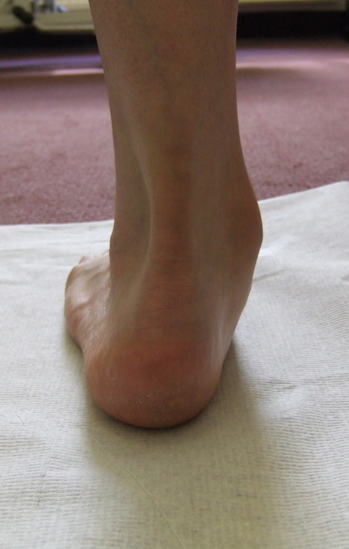
As the tendon stretches pain on the inside of the ankle may settle, but ‘dysfunction’ becomes a problem, walking becomes more of an effort due to weakness and the foot lacks ‘push-off’, sportsmen find it difficult to run and jump. People often notice a change in the shape of the foot, describing flattening or turning out of the foot. Pain develops over the outside of the ankle as the foot turns more outwards.
How is Tibialis Posterior Tendinosis diagnosed?
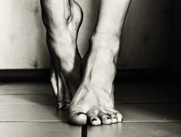
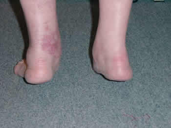
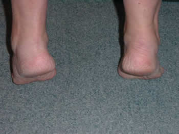
The diagnosis of tibialis posterior tendinosis is usually clinical, you will be carefully examined to identify the location of your pain, deformity of the foot, flexibility and function. You will be asked to attempt to stand on the tiptoes of both and then one foot. Whereas in a normal flat foot if one stands on tip-toes, the foot forms an arch as the heel swings inwards, in an adult acquired flat foot the arch doesn’t reconstitute and the heel does not turn inwards, it also becomes very difficult for one to stand on the tiptoes of one foot.
An x-ray is usually performed to identify any degenerative change within the hind-foot joints. Occasionally an MRI or Ultrasound scan may be needed.
How is Tibialis Posterior Tendinosis treated?
Early tibialis posterior tendinosis, where the foot is still flexible may be treated with special orthoses (insoles) that support the arch and hold the heel in a corrected position. Physiotherapy to strengthen the tendon and stretch the calf muscles should also be performed.
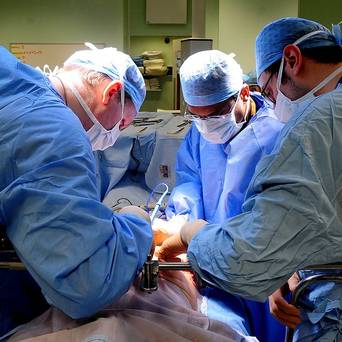
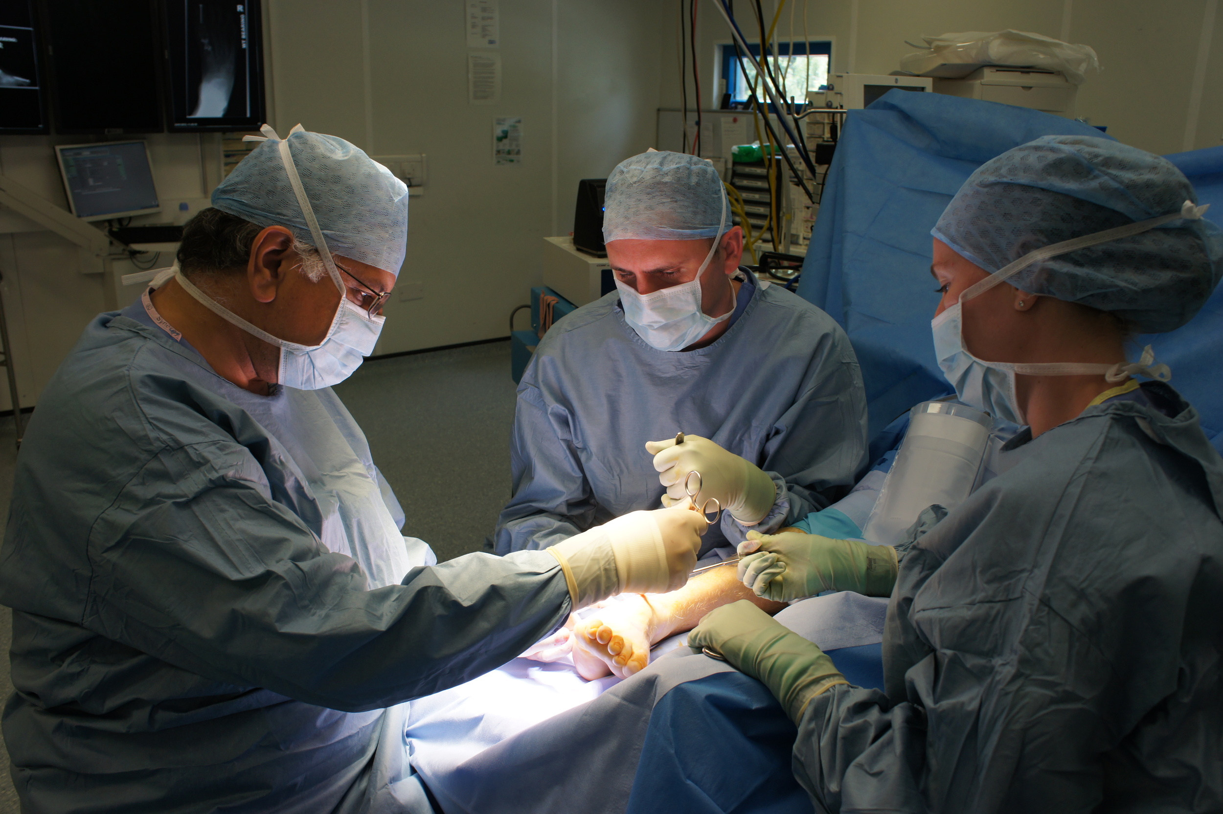
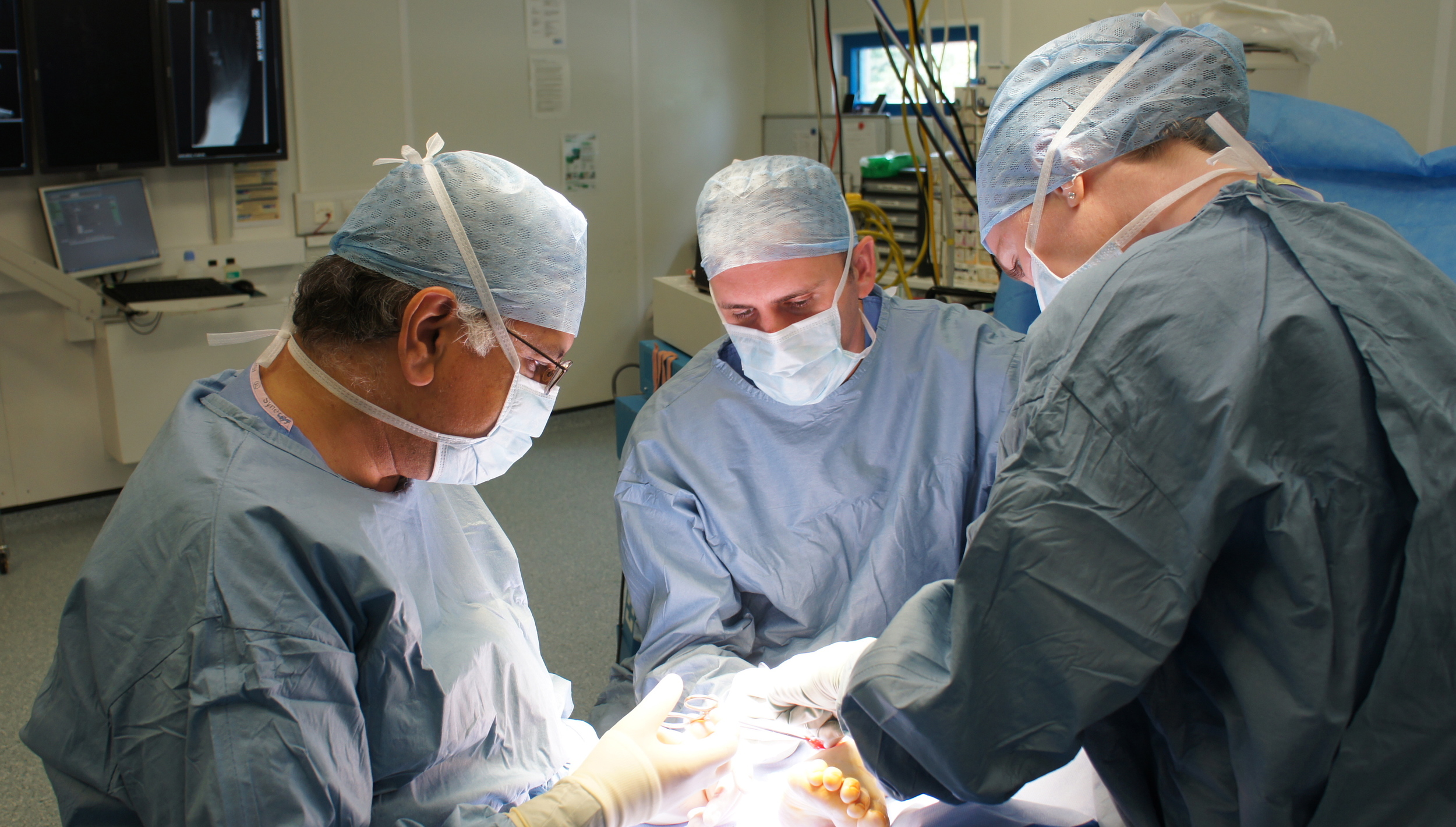
A surgical photograph of a diseased tibialis posterior tendon.
If simple treatments fails surgery may be required.
Flexible foot: A tendon transfer is used to replace the failed tibialis posterior tendon. The tendons that we use are either the tendon to the toes (FDL) or part of the strong tendon over the front of the shin (Tibialis Anterior) and these are fixed to the navicular bone using stitches or special screws, in the same place that the tibialis posterior tendon was attached.
Part of this procedure is to re-align the heel so that it lies underneath the body, as it should, and helps take the strain off the new transferred tendon. The back of the heel bone (calcaneum) is cut using a saw which allows it to be shifted across inwardly before being fixed with a screw.
The aim of this surgery is to correct the foot position, relieve pain and maintain the movement of the foot.
After this surgery you remain in a below knee plaster for 6-8 weeks, initially the foot looks to be turned inwards, the plaster needs to be changed every 2 weeks in order to bring it around into a more normal position. click here for link to tibialis posterior information sheet PDF. After surgery you will require intensive physiotherapy to strengthen the new tendon and teach it its new role and maintain movement in your hindfoot. On the whole you will continue to improve over many months:

3 months: Fair
6 months: Good
12 months: 'Right'
Stiff Foot: In longer standing cases the joints of the heel are stiff and painful, which means that a tendon transfer is not possible. In these cases a ‘triple arthrodesis’ may be required. This is an operation that corrects the deformity of the foot by fixing the joints solidly in place and allowing them to fuse (arthrodesis). This operation although providing good pain relief, leaves the hindfoot stiff, limiting the sideways movement of the heel, the ankle joint is not affected.
After a triple fusion you will require to remain in a below knee plaster of Paris for twelve weeks. Click here for link to hindfoot arthrodesis PDF.
Be sociable..share!